Stereology is a tool for obtaining unbiased estimates of object numbers and/or sizes within three-dimensional regions of interest. For biological samples, quantitative measurements are potentially more objective and accurate when compared to traditional semi-quantitative grading of findings by pathologists (i.e., scoring of morphologic changes as minimal, mild, moderate, or severe). Although both approaches require high resolution cameras and specialized software, stereology differs from conventional two-dimensional image analysis methodology in several respects. First, stereology is usually performed using projected live slide images or image stacks, as opposed to individual digital photomicrographs. Second, stereological evaluations are conducted in three planes (x, y, and z) as opposed to two (x, y), which allows for more accurate estimation of measurements such as volume, surface area, and connectivity, in addition to object counts and length determinations. Unlike techniques used decades ago, modern design-based stereology accomplishes this without resorting to assumptions concerning the size, shape, or spatial distribution of objects throughout the region of interest. Finally, stereological methodology typically incorporates stringent randomization measures that are used to mitigate sampling and observational bias; similar measures could (and should) be included in image analysis projects but are frequently lacking.
Preparation of Thick (40 µM) Free-Floating Mouse Brain Sections for Stereological Assessment
The decision to use a stereological approach versus other methodologies typically depends on a number of factors that include sample type suitability, the magnitude of the expected morphological changes, cost, turnaround time, and the anticipated acceptance of study findings by the target audience. For example, stereology is generally considered to be the gold standard for quantitative assessments of the central and peripheral nervous systems and can support data from nonclinical biomarker qualification studies (Food and Drug Administration, 2016). Therefore, journal reviewers and regulatory agencies routinely expect to see stereological data as the output for those particular types of studies. However, the stereological approach is applicable to a variety of organ systems and tissue types. Examples relevant to toxicologic pathology include: ovarian or testicular gametocyte counts; volume measurements of emphysematous lung bullae, thyroid follicles, or renal glomeruli; area and volume measurements of neuron subtypes in ganglia, length and density measurements of intraepidermal nerve fibers, determinations of mitochondrial numbers and sizes in transmission electron micrographs; and assessments of the degree of angiogenesis inhibition in tumor explants.
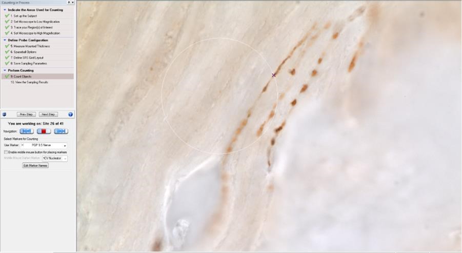
Space Balls Probe for Length Estimation of PGP9.5 Immunoreactive Nerve Fibers
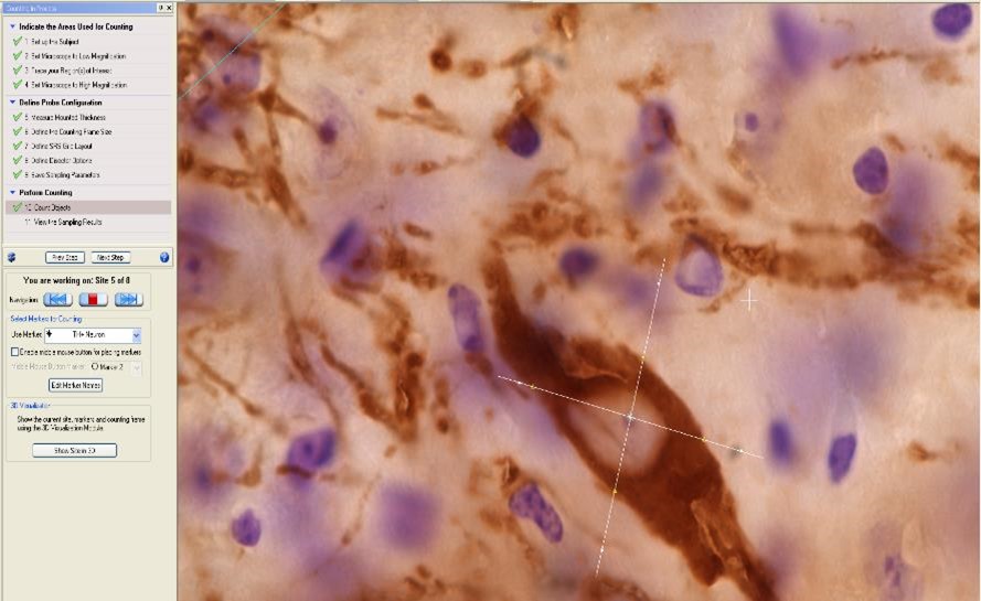
Nucleator Probe for Volume Estimation of Tyrosine Hydroxylase Immunoreactive Neurons
Tissue sample quality and uniformity are often the most critical components of any stereology study. Consequently, experiments that were not designed to be stereology experiments a priori are commonly plagued by problems of data inconsistency; alternatively, the incorporation of method development work and pilot studies are recipes for successful stereology projects. EPL has been conducting stereological investigations in an industry environment continuously for the past ten years.
REFERENCES
Food and Drug Administration. (2016, May). Considerations for Use of Histopathology and Its Associated Methodologies to Support Biomarker Qualification – Guidance for Industry. Silver Spring, Maryland, U.S. Retrieved from https://www.fda.gov/ucm/groups/fdagov-public/@fdagov-drugs-gen/documents/document/ucm285297.pdf